Tiny Fighters: Dipping my Toes
Hi everyone! Welcome back to my blog!
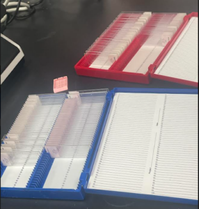
During my time working at the lab at the University of Arizona, I was fortunate enough to have the opportunity to work closely with Dr. Halpern. We spent a considerable amount of time observing and analyzing NEC stain samples in both infant and mouse/rat liver gut tissue. This allowed us to study the histology of the samples in-depth and gain a better understanding of the pathogenesis. Through the use of advanced technology and specialized software, I was able to learn about the detection of NEC. I discovered that this is a crucial aspect of the disease, as it enables healthcare professionals to intervene, diagnose, and prevent further complications. Moreover, I was able to learn about the 4-point scale that is used to measure the severity of NEC. This scale allowed us to express the severity of the disease clearly and concisely and provided valuable insights into how the disease progresses over time.
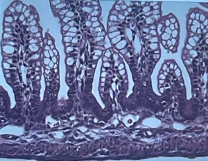
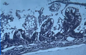
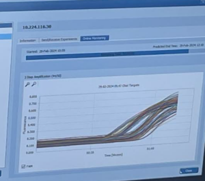
We observed two samples that were stained with H&E to show the difference between healthy and unhealthy liver tissue. The first image showed thick and healthy villi and stem cells that form the structures. However, in the second image, the villi was necrotized, as can be seen by the distortion near the top, and in the enterocytes, lumen, and mucosae. We then proceeded to learn about histology and the research process behind medical diagnosis and forensic applications. After that, we moved on to practical methods for detecting NEC. Our approach involved loading 96 wells of 7 different proteins that have shown unusual expression in cases of NEC under gel electrophoresis. Furthermore, we have identified those 7 several proteins that can be used to detect NEC. This enables us to visualize the PCR reaction. We then collected the data and performed a T-test under a p-level of 0.95 to determine if there was any statistical significance. The results showed that the seven proteins exhibited statistical significance and could be detected under fluorescence along with their association with ASBT. Our study aims to provide a better understanding of the histological differences between healthy and unhealthy liver tissue. The images below showcase the PCR reaction and the T-test calculations, which support the validity of prior hypotheses.
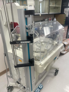
I was given a sneak peek into what our next meeting might entail. During the meeting, we discussed an incubator that is typically used in neonatal intensive care units (NICUs) to create a safe and controlled environment for premature infants to develop their vital organs. The NICU incubator was modified for research purposes. In this case, mice were placed in the incubator instead of babies. Since mice tend to hover and snuggle against each other for skin-to-skin contact in their litter, they are usually placed in small plastic cups with bedding to separate the pups, mimicking the conditions a baby would be placed in. This allows for accurate research on necrotizing enterocolitis (NEC) by collecting data from mice.
As I explored the lab, I couldn’t help but hope that on my next visit, I would get the opportunity to work with mice. There’s so much to learn, and I’m eager to take it all in! Thank you for reading!

