Tiny Fighters: Critical Analysis
Hi everyone and welcome back to my blog!
In my last blog post, I shared my experience of working in the lab with Dr. Halpern and research assistant Christine. I got the chance to observe and learn about the histology of NEC, PCR, and gel electrophoresis. We studied the different proteins involved in NEC using a T-test to determine their statistical significance. During our discussions, we also touched upon the fluorescence of Apical Sodium-Dependent Bile Acid Transporter (ASBT) and Western blotting for NEC. Exploring these topics further would help to understand the biological mechanisms that contribute to NEC. Although I have previously written about bile acids and their role in this condition, ASBT is a crucial factor that plays a significant role in NEC development in premature infants.
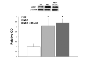
The figure above illustrates western blotting and data from ASBT expression in rat and mouse models of NEC. Because SC -435 is a competitive inhibitor of ASBT, the data shows how ASBT protein levels in the NEC + SC – 435 group were not significantly different from ASBT protein levels in the NEC group that were not given the inhibitor.
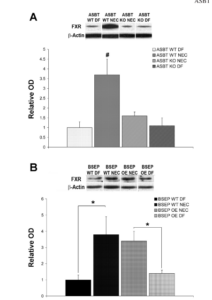
In this image, the data evaluates Farsenoid X receptor (FXR) protein expression in experimental NEC. ASBT expression is controlled by a negative-feedback regulatory loop, where hydrophobic () bile acids (CDCA and CA) can transcriptionally regulate their enterohepatic circulation through the activation of FXR. Enterohepatic circulation is where bile acids are moved from the liver to the small intestine and back to the liver. FXR protein was significantly higher only in the ASBT WT NEC (ASBT wild-type NEC) than in the other three groups. However, FXR protein was elevated in BSEP WT NEC (Bile salt export pump wild-type NEC) and BSEP OE NEC (Bile salt export pump overexpressed NEC). FXR activation should inhibit ASBT and decrease bile absorption but the data in this image proves to contradict this. These images explain more about how ASBT, FXR, and bile acids overlap and influence each other to form NEC.
The three images above with fluorescence observe the stains of anti-ASBT under three different severities of NEC. The green fluorescence represents ASBT and correlates to whether that infant has NEC and a rough approximation of severity.
NEC is a complex condition that involves various biological mechanisms such as protein expression, pumps, inhibitors, and bile acids. A deeper understanding of these mechanisms can help identify the factors responsible for NEC and how they regulate each other to cause the condition. I will be heading back to the lab soon to learn more about these mechanisms. Furthermore, I am hopeful to get hands-on experience in dealing with mice in the incubator. Thank you for reading!
https://drive.google.com/file/d/1MwynlACHfj4aAAVEAyPES-B8taf6wOrM/view?usp=sharing


