Week 3: Finishing Lab Work, Collecting Results…And an Introduction to Beekeeping!
Riya d -
**To stay consistent with my peers, this blog update will discuss my experiences during the third week of my internship (February 17 to February 21)**
Hello everyone, welcome to Week 3 of my project!
Our goal this week was to finish the experiments for my project so I could spend some time assisting with other work in the lab. We started by trying to score the slow-growing bacteria inhibition plates from the past week. Unfortunately, we had to redo many problematic plates due to unscorable, uneven zones of inhibition and sparse growth.
While awaiting results from the redo plates, I finally had some time this week to visit the bee yard! Initially, the plan was for me to harvest royal jelly from the hives and make inhibition plates comparing the fresh jelly to the frozen jelly that the lab typically uses in their experiments. However, we quickly realized this wasn’t going to be feasible. We had already been apprehensive about collecting royal jelly this season, as it involves de-queening a hive (so that it would produce a lot of royal jelly in its attempts to rear a new queen that we could then harvest), and hives are not very active and strong immediately after the cold winter months. Instead, we had considered harvesting worker jelly, which is very similar to royal jelly and found in the cells containing brood (larvae are fed this).
However, when we tried to collect worker jelly from a hive frame, we discovered that the hive was not only producing very little new brood but was also sick (see Figure 1). Many larvae, according to my mentors, seemed to be exhibiting symptoms of European Foulbrood. Consequently, there was insufficient worker jelly for the experiments, and we decided to cancel the fresh versus frozen royal/worker jelly inhibition assays.
 |
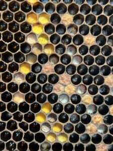 |
Figure 1. The hive frame set on a frame stand (left). On the right: Close up of the hive frame. Bright yellow cells are filled with pollen, dark ones with a brown tinge contain honey, white cells contain curled up larvae in worker jelly, and the cells that appear to be closed are “capped cells” containing larva that are developing into pupa. Other dark cells that may seem empty actually contain tiny, rod shaped eggs, visible when zoomed in.
Instead, I had the opportunity to get a taste of beekeeping! After first ensuring I was essentially “sting proof” (a shirt and jacket under a full bee suit, a hat plus a veil, double gloves, and boots), I learned how to properly handle the hive frames and got to assist my mentor with doing some “sugar shakes”. Sugar shakes are used to check for the presence of varroa mites in hives, a common parasite that affects honeybees.1 To do this, we gently brushed about 300 bees from a hive frame into a mason jar, added powdered sugar, and shook the jar to coat the bees in the sugar. We then turned the jar upside down, allowing the sugar and also any mites to fall out of holes in the lid onto a paper towel. After spraying the towel with water to dissolve the sugar, we could easily spot the mites. Depending on how many mites/300 bees we count, we treat the hive accordingly (this lab chooses to use formic acid for mite control).
Between finishing up my essential oil experiments and spending time with the bees, this week was buzzing with excitement! Next week, I will score the redo plates from this week (hopefully with no further issues!) and start analyzing my data.
See you then!
References
- Carroll, M. J., & Brown, N. J. (2024). Varroa mite removal from whole honey bee colonies by powdered sugar dusting is enhanced by crowding and mechanical agitation of treated workers. Journal of Apicultural Research, 63(4), 637–647. https://doi.org/10.1080/00218839.2024.2361959
Comments:
All viewpoints are welcome but profane, threatening, disrespectful, or harassing comments will not be tolerated and are subject to moderation up to, and including, full deletion.