
Microglia Activation Post Injury
Pariza F -
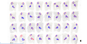
This week I completed brain imaging as well as the calculations needed for analysis. Every individual brain slide came from a random rat that had experienced a traumatic brain injury. Each slide makes up a different portion of the brain. Therefore, when you put the first 32 brain slides together, they should make a 3D version of a rat brain. Using this method, we can see how a traumatic brain injury affects every aspect of the brain in at least two sets of brains (64 brain slides).
For each brain slide, we have picked three sections to analyze by counting the amount of microglia in each which are:
VPM: primary relay center for sensory information in the face, head, and oral cavity
RTN: regulates neurons for respiratory purposes such as changes in carbon dioxide levels in the blood
S1BF: processing tactile information; for rodents, whiskers are affected since they are used to pick up movements
We are looking to detect differences in microglial activation at seven days post-injury(DPI) while taking sex differences into account. Taking both of these things into our analysis, we have made two hypotheses:
Hypothesis 1: Diffuse axonal injury leads to chronic neurodegeneration responsible for chronic
activation of microglia.
Hypothesis 2: Previous literature indicates a sex-dependent trajectory in the level of microglial activation
and the morphological profiles after a focal cortical injury induced by controlled cortical impact
(Villapol). Sex differences were predominantly early post-injury. Microglial activation out to 6 months
post-injury has not been quantified in thalamic nuclei and cortex after diffuse axonal injury by midline
fluid percussion.
With the amount of microglia activation present in the S1BF region which affects whisker sensation, I believe there may be neurobehavior relevance in that area. Yet, other studies show a delayed neuroinflammatory response in females so we must do further research on this criterion as we suspect that female data will be distinct from males.
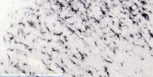
The images show a close-up of active microglia cells and the brain slides. When you put all the brain slides together, they should make an entire rat brain which is pretty cool!
Comments:
All viewpoints are welcome but profane, threatening, disrespectful, or harassing comments will not be tolerated and are subject to moderation up to, and including, full deletion.