Chromosomes, Cameras, and Conundrums
Tisya o -
“Centered, in focus, vivid colors…that’s a perfect photo!”, I thought to myself. In my quest to learn more about the new northern gelada species, I took pictures of wild gelada monkey chromosomes this week.
This painstaking process begins with cultivating cell lines in a university in Nevada, as my advisor Brooklynn explained. These cells are then shipped to ASU where my advisor treats them with g-banding. To do this, she arrests these cells in metaphase (the second phase of Mitosis where the chromosomes align in the center of the cell), treats them with a hypotonic solution, places them onto a microscope slide, treats them with trypsin, and, finally, stains the cells with Giemsa stain. The final result is a beautiful, rich blue explosion of gelada chromosomes with vibrant light and dark bands.
However, the real challenge comes in documenting these little masterpieces. Like most natural wonders, a simple picture cannot do justice to all the fine, exquisite details, but still, I had to try. I clumsily picked up the microscope slide and placed it on the stage, but to my surprise, I couldn’t see any chromosomes or anything at all. After my advisor informed me I was looking at the slide’s label and readjusted my microscope, a bunch of tiny and even tinier blue dots filled my vision. These tiny blue dots were the entire basis of my project: thousands of gelada chromosomes. To capture the best quality photo I used a process called oil immersion. After plugging in my laptop to the microscope to capture an image of what was on the slide, I placed a drop of oil on the microscope slide and used the 100x magnification lens to capture the clearest images of these chromosomes. Even the tiniest movement would unfocus the entire microscope and completely compromise my image, so I had to take the utmost care and precision. After two hours, I emerged (somewhat) successful with five images of different chromosome spreads.

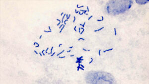
I am currently in the process of counting the number of chromosomes in each image; I hope to consistently see 44 chromosomes in each image as that would align with the recent discovery of a 2n=44 chromosome karyotype in a sample of northern geladas. With every successful chromosome image, my case for the 2n=44 karyotype in northern geladas and the formation of a new gelada species becomes even more concrete. Alongside this process, I am working on filtering raw data to eventually create a PRDM9 graph (a visualization of where the PRDM9 protein binds to DNA). By understanding where PRDM9 binds, we can see where it is more likely that meiotic recombination is occurring. Recombination is one of the biggest contributors to chromosomal evolution, so by exploring PRDM9 and recombination hotspots, I hope to understand more about chromosomal evolution in geladas and the fission of chromosome 7 in northern geladas.
Materials Referenced;
- Chiou, K. L., Janiak, M. C., Schneider-Crease, I. A., Sen, S., Ayele, F., Chuma, I. S., Knauf, S., Lemma, A., Signore, A. V., D’Ippolito, A. M., Abebe, B., Haile, A. A., Kebede, F., Fashing, P. J., Nguyen, N., McCann, C., Houck, M. L., Wall, J. D., Burrell, A. S., Bergey, C. M., … Snyder-Mackler, N. (2022). Genomic signatures of high-altitude adaptation and chromosomal polymorphism in geladas. Nature ecology & evolution, 6(5), 630–643. https://doi.org/10.1038/s41559-022-01703-4
- Guarracino, A., Buonaiuto, S., de Lima, L.G. et al. Recombination between heterologous human acrocentric chromosomes. Nature 617, 335–343 (2023). https://doi.org/10.1038/s41586-023-05976-y
- Mayo Clinic. (n.d.). Routine cytogenetic analysis. Mayo Clinic. Retrieved February 23, 2025, from https://www.mayo.edu/research/core-resources/cytogenetics-core/services/routine-cytogenetic-analysis
- Pritchard, J. (2024). Human genome variation and why it matters. In An owner’s guide to the human genome: An introduction to human population genetics, variation, and disease (Chapter 1.3). Stanford University.
Comments:
All viewpoints are welcome but profane, threatening, disrespectful, or harassing comments will not be tolerated and are subject to moderation up to, and including, full deletion.